
No products in the cart.


Virus like particles (VLP) as vaccine
Virus-like particles (VLP) Platforms for immunogens, vaccines and drug carriers
Antibody-drug Conjugate (ADC): Pre-made ADC benchmark, MOA, Production and QC
Neutralizing antibodies of virus (SARS2, HIV, HBV, Rabies, RSV, Ebola, Influenza)
Immunoglobulin Fc receptors for Fc&Fc Receptor binding assay
ILIBRA-HuEasy Monoclonal antibody (mab) humanization service (fully humanized ab)
Single domain antibody (Nanobody)
SOCAIL MEDIA

The repeated units of protein monomer on the VLP surface are a strong immunogen. Compared to subunit peptides or proteins, epitopes on the VLPs surface have the natural structure to stimulate the B- and T-cell immune response. Moreover, repetitive epitope on the surface of VLPs stimulate the strong adaptive immunity without the help of adjuvants.
VLP-based vaccines are safer than inactivated or attenuated vaccines. incomplete inactivation and attenuation might reverse the virus activity and cause infection, but the lack of viral genome in VLP could not cause infection at any time point. In addition, the epitope can be incorporated onto the VLP surface through different in-vivo and in-vitro techniques as mentioned above. For example, Qb bacteriophage virus-like particles (Qb-VLPs) have been used as a vaccine for the non-immunogenic week immunogenic antigens.
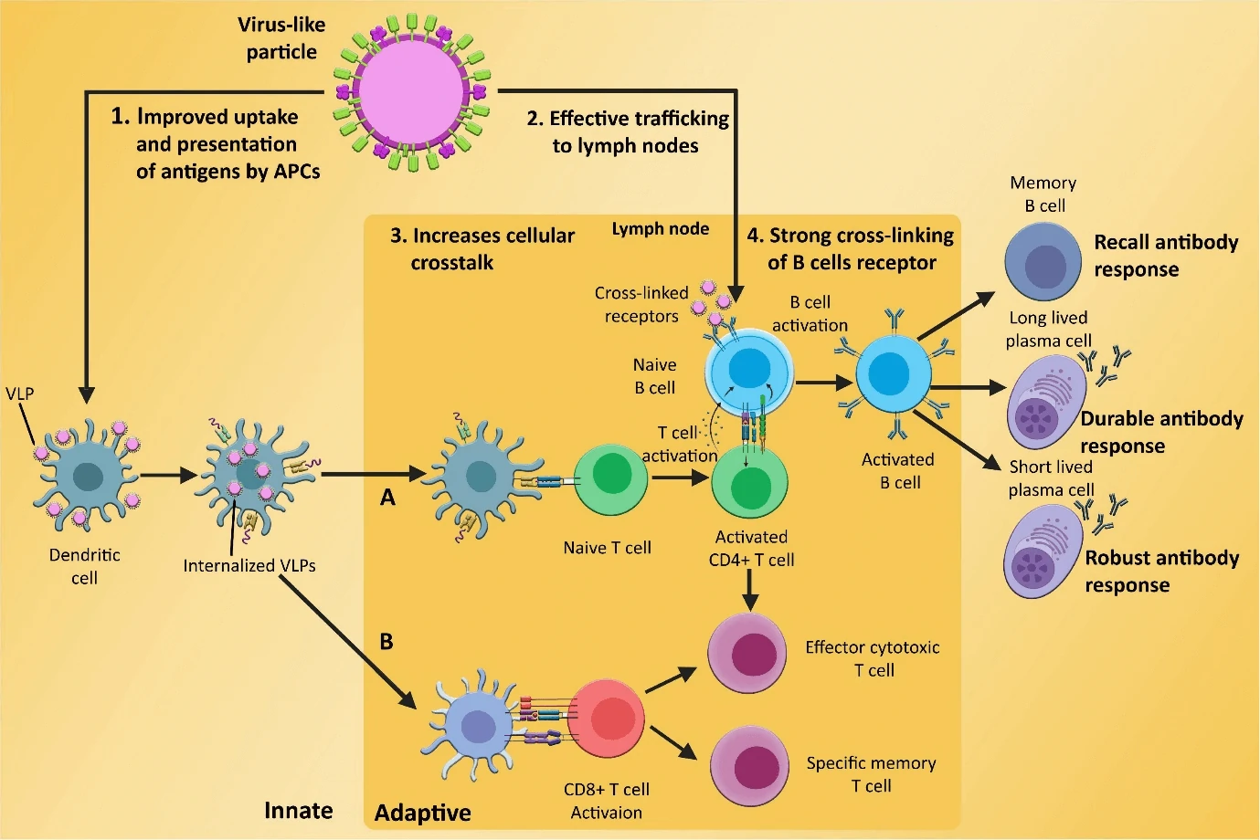
In response to the virus like particle vaccine, the markers such as CD40, CD80, and CD86, on the surface of the dendritic cells (DCs) are expressed and activate the DCs. The activated DCs bind with VLPs through the pattern recognition receptors (PRRs) such as Toll-like receptors (TLR2).
Later, VLPs are internalized to the cytosol of DCs and presented to cytotoxic T cells and helper T cells through MHC class I and class II molecules, respectively. Cytotoxic T cells (red triangles) kill pathogens/tumor cells by discharging cytotoxic proteins T helper cells activate cytotoxic T cells by releasing interleukin-2 (IL-2) and interferon gamma (IFNγ). On the other hand, T helper2 cells induce B cells to secrete antibodies (Hasnat et al. 2022).
View the Knowledge base of virus like particles (VLP):
– Virus-like particles (VLP) Platforms for Developing immunogens, vaccines and drug carriers
– What is virus like particles (VLP)?
– GM VLPxTM Virus-like particles development platform
– Virus like particles (VLP) Expression platforms
– Virus like particles (VLP) as immunogens/antigens of transmembrane protein (TM)
– Virus like particles (VLP) for drug delivery
– Virus like particles (VLP) as vaccine
– Virus like particles (VLP) for drug discovery
